Plant Cells: Comparing Monocot and Dicot Stems

This simple, step-by-step lab activity provides a sense of realism and participation that commercially prepared slides or illustrations can't possibly duplicate. Students prepare slides using pre-cut monocot and dicot plant cross sections and macerated stem tissue. After staining and mounting the samples, students then view their slides through a microscope. Students use clearly labeled diagrams and a dichotomous to help guide them in locating and identifying a variety of different types of cells.
Details at a Glance
- 1-2 Days | 1-2 ~50 minute class periods
- 1 Activity
- Accommodates 2 classes, each with 15 groups of 2 students
- Consumables service 60 students
- Meets our criteria for supporting literacy
- Includes digital resources
- Requires a water source
Scientific Concepts
•Preparation, staining, and mounting of microscope slides
•Observing cells under a microscope.
•Utilizing diagrams and dichotomous keys for identification.
•Comparing the stem structure of monocots and dicots.
KEY VOCABULARY: dicot, monocot, phloem, pith, vascular bundle, xylem
Guides & Student Sheets
Our kits and modules provide you with everything you need so you can open, review, and teach the material confidently the next day.
- Comprehensive Teacher Guide with background information, detailed instruction, example data and answers
- Student Sheets with age appropriate background information, full procedure(s), and analysis items
- Materials necessary for the investigation (beyond common classroom items)
- Safety Data Sheets
Kit Components
- 90 Microscope slides
- 90 Cover slips
- 30 Tissue transfer brushes
- 4 Drop controlled bottles of Differential Stain
- 1 Drop controlled bottle of Glycerine
- 1 Vial of Dicot Cross-sections
- 1 Vial of Macerated Stem Tissues
- 1 Vial of Monocot Cross-sections
- 30 Student Guides
- 1 Teachers Guide with MSDS
- Not included and needed for instruction: Class set of safety eyewear, class set of microscopes.
Need to order refills for this Kit?
Order RefillsRelated Products
-
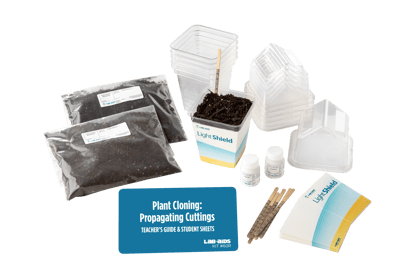
 Kit #60RPlant Cloning: Propagating Cuttings
This life science kit provides students with specially designed materials that make it easy to clone plants through the process known as asexual plant propagation. This activity requires no special facilities for plant propagation because the Lab-Aids® Greenhouses automatically control many environmental conditions. The Lab-Aids® Greenhouse was designed for small, controlled investigations...
$163.40 View Details -
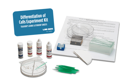
Kit #63Cells Experiment
Cell Differentiation is a process by which development of a living organism is achieved. Through a series of progressive changes, a generalized cell transforms into a specialized cell. The more specialized the cell becomes; the less likely it is to divide. After the seeds germinate, students prepare slides, then watch living material differentiate before...
$101.10 View Details -
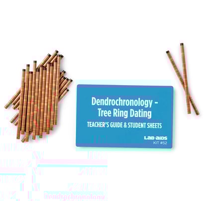
Kit #52Tree Ring Dating
This activity provides a basic knowledge of the principle of tree ring dating by using simulated core samples. Students first determine the age of a “tree cut down in 2019” then extend their understanding of tree ring correlation by determining the age of a “tree cut down” decades ago.
$80.80 View Details -
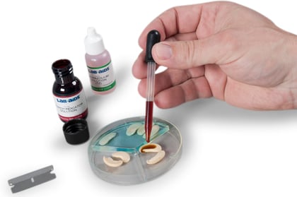
Kit #66Seed Staining
In this engaging and fun kit, students learn how to differentiate between plant food stored as a simple sugar and food stored as a starch. Using a step-by-step procedure, students first dissect the seeds, and then treat them with reagents. This helps to determine the quantity and location of the sugar and starch. The...
$102.30 View Details -
Kit #65Seed Structure
An excellent activity for middle school through college, this kit focuses on the structure of dicot and monocot seeds. Students first examine and dissect two representative types of seeds - dicotyledons and monocotyledons. After diagramming and labeling seed structures, they study the action of enzymes in the germination process and the fate of each...
$154.60 View Details